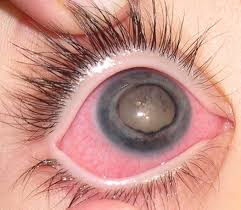
Coats disease, also known as exudative retinitis, is a very rare eye condition that affects the smaller blood vessels (capillaries) found in the retina.
The retina is the light-sensitive layer that lines the inside of your eye. Coats disease can make these blood vessels weak and grow incorrectly, causing them to leak fluid and blood under the retina. This means that the cells of the retina can’t work properly, and this can cause sight to be affected.
Coats disease affects males more than females. It’s usually diagnosed by the time a person is 20, but in most cases, it’s first detected in childhood before the age of 10. Adults can also be affected, and some people, often middle-aged males, can have a milder form of the condition.
Coats disease only affects the health of the eye so people with the condition are otherwise generally healthy.
What causes Coats disease?
The cause of Coats disease isn’t fully known (the medical term for an unknown cause is idiopathic) and there are no known risk factors that make it more likely to develop. Coats disease is not an inherited condition meaning that it is not passed on within families. As the condition is rare, it’s extremely unlikely for it to affect more than one child or person in the same family.
It has been suggested that for some people, Coats disease may be related to a mutation (fault) in the NDP gene which develops after conception, meaning that the condition happened by chance and was not inherited from their parents.
How does Coats disease affect the eye?
Coats disease affects the smaller blood vessels (capillaries) in the retina. Retinal capillaries are important in supplying the retina with blood which carries nutrients and oxygen to its cells, so that they work correctly. The cells of the retina need to remain healthy for you to be able to see clearly.
Coats disease causes retinal capillaries to develop incorrectly. They become wider (dilated) and twisted, which make them more noticeable when the inside of the eye is examined. The medical term for these changes is telangiectasia.
As well as becoming dilated, the retinal capillaries also become weak and leaky. This causes some of the fluid from the blood to leak out of the vessels and into the retina. This fluid builds up in the retina and causes it to become swollen. Eventually, exudates (proteins and lipids which have leaked out of the vessels) build up underneath the retina, causing a yellow appearance in the affected area.
Where there are areas of exudates and telangiectasia, the retina won’t be able to work properly. This in turn may affect your sight.
The stages of Coats disease
Coats disease has different stages depending on how it is affecting the retinal capillaries and retina:
Stage 1: Telangiectasia (dilated and twisted capillaries) which cause minimal change to the retina and to vision.
Stage 2: Telangiectasia and exudates which cause increased changes to the retina and, if uncontrolled, may lead to changes in vision.
Stage 3: Large areas of swelling with retinal detachment. Vision is likely to be poor at this stage.
Stage 4: Complete retinal detachment and glaucoma (raised eye pressure). These complications can be treated, but sight is still likely to be very poor.
Stage 5: The eye has no sight, and there is no treatment that can improve sight. In most cases the eye isn’t painful, but if pain develops, treatment may be needed for this, and to try to preserve the eye.
How does Coats disease affect sight?
For most people, Coats disease only affects one eye, so many with the condition will have good sight in the other eye.
Coats disease is a condition which can get worse, affecting more of the retina over time, but it can stop getting worse on its own, so that not everyone progresses to stage 5. It’s not known why the condition can affect some people more than others, making it very difficult to predict how it may progress over time. Generally, the outcomes for sight are poorer in younger children who are at a more advanced stage of the condition when they are first diagnosed. The retinal changes tend to progress more quickly in younger children, especially those under three years old. Those changes are more difficult to control and are more likely to lead to a greater long-term reduction in their vision. When the condition develops in older children and young adults, it can cause much milder retinal changes which progress more slowly, so that their vision is less affected over time.
In the early stages of Coats disease, a person’s peripheral vision is most likely to be affected. If the condition progresses, there will be a greater loss of vision in this eye. If Coats disease affects the macula, then a person’s central, detailed vision will be reduced.
Will I notice any signs of Coats disease?
Often, an eye examination is the only way to tell if your child has an eye condition. Most children with Coats disease don’t have any symptoms – the eye doesn’t look unusual, and it isn’t painful or red. Many children have their vision screened when they start school at the age of four or five. However, this does not happen in all areas of the country. If you’re concerned about how well your child can see, or their vision is not screened at school, taking them to an optometrist (optician) will mean their eyes can be fully examined and their sight checked.
Sometimes, you may notice that your child’s eye has an odd appearance in photographs, particularly where flash photography is used. Normally, flash photography causes a red-looking pupil but an eye with Coats disease may have a pupil that looks white or pale yellow instead, which some people describe as a glow in the eye. A white pupil is known medically as leukocoria. If you notice that your child has the appearance of leukocoria at any time, it’s important that their eyes are examined urgently by an optometrist or ophthalmologist (hospital eye doctor), because as well as Coats disease, there are other serious eye conditions that can give a similar appearance which need to be ruled out.
Some children who have Coats disease may develop a squint. This is sometimes described as having a “turn in the eye”. Having a squint means that their affected eye doesn’t look in the same direction as the other eye.
However, the only way to diagnose Coats disease is by examination of the back of the eye. If an optometrist has examined the back of your child’s eye during a routine sight test and believes they may have Coats disease, they will refer them to the eye hospital to be seen by an ophthalmologist.
How is Coats disease treated?
The aim of treatment is to stabilise any changes that are already on the retina and to prevent sight from getting any worse.
In the early stages of Coats disease, vision may not be affected, and for some people, Coats disease won’t develop any further than stage 1. If the condition is very mild, treatment may not be needed straight away, and the ophthalmologist may decide to monitor the eyes at regular appointments instead.
However, where there are a lot of changes to the retina, even when the vision remains good, the ophthalmologist may decide to carry out some treatment to help prevent a deterioration in sight.
The treatment offered will depend on what areas of the retina have been affected and by how much. Coats disease can be treated using laser photocoagulation and cryotherapy (freezing treatment). The aim of both these treatments is to help seal up the retinal capillaries to stop them from leaking further. Most children would have these treatments carried out under a general anaesthetic.
In more advanced stages of Coats disease, where the retinal capillaries have leaked a lot and the swelling has caused the retina to detach from the back of the eye, the treatment will be aimed at re-attaching the retina. The type of treatment may vary depending upon how large the detachment is and how long the retina has been detached for but may include surgery to re-attach the retina.
Occasionally, in certain circumstances, injections may be given into the eye. These treatments may include either anti-VEGF medication or steroids.
Does Coats disease lead to any other complications?
As well as retinal detachment, there may be other complications for someone with Coats disease which also require treatment, such as uveitis, the development of a cataract, or glaucoma. Cataract and glaucoma are also side effects that can develop from having steroid treatment.
There are, unfortunately, some people for whom treatment is not successful because their eye doesn’t respond to any of the treatments they receive. In these cases, it’s possible that they may lose all the sight in their eye. Usually, their eye won’t become uncomfortable. However, very rarely, glaucoma develops which cannot be controlled, and this can cause the eye to become very painful. If the eye pressure remains too high and causes continual and intense pain, the ophthalmologist may suggest that removing the eye and replacing it with an artificial one is the best next step to take, but this is always a last resort and is not suggested very often.
Most people with Coats disease do not experience this type of complication and so they will not need to have their eye removed.
How does someone manage with sight in only one eye?
Coats disease usually only affects one eye while the other eye may have good sight. When someone has good sight in one eye, and poor or no sight in the other, they have what is known as monocular vision.
Children tend to adapt very well to using their better eye. This doesn’t mean that they will be overusing or damaging their better eye as this adaptation is a natural process. It’s unusual for children with good vision in one eye to need additional support in their education. People with monocular vision are not considered partially sighted.
Having monocular vision can affect depth perception and hand-eye coordination. With some tasks, your child may appear clumsy and uncoordinated at first, for example when they throw and catch a ball. However, this generally improves as your child gets older and adapts to being monocular.
Even with good vision in their better eye, someone with monocular vision doesn’t have as much peripheral vision as someone with two eyes. With time they will adapt to this without realising by turning their head more to see things around them. Other people can help by sitting or approaching someone with monocular vision on the same side as their good eye, to make it easier for them to be seen.
It’s important for people who have good sight in only one eye to have regular eye tests with an optometrist so that the health of their good eye can be monitored. Your optometrist should let you know how often you need to have your eyes examined.
For people who have sight in only one eye, it may be a good idea to consider the use of protective eyewear or sports goggles for certain sports or activities to help prevent injury to the good eye. You can speak to a dispensing optician at your local optician’s practice for more advice about this. Dispensing opticians are qualified in the dispensing and fitting of spectacles and can give professional advice about suitable frames and lenses.
People with monocular vision are still able to drive a private vehicle (Group 1) as long as the vision in their better eye is unaffected by any other eye conditions, and it meets the visual requirements for driving. However, monocular vision would mean you couldn’t hold a heavy good vehicle (HGV) or public service vehicle (PSV) licence (Group 2). Some professions, such as being a pilot, police officer or certain roles within the Armed Forces require a specific level of vision to be reached in both eyes. Keeping this in mind can help with planning a career choice for the future.
