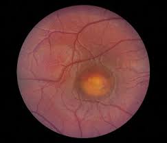
Best disease affects the macula, which is the central part of your retina at the back of your eye. which you use when reading, writing, or watching TV. There is no current treatment for Best disease at present, although research into is on-going in the area of gene therapy is on-going, which may lead to a treatment in the future.
This page contains a summary of our information on Best disease. To read our full information, download our factsheet:
What is Best disease?
Best disease is a type of macular dystrophy and is also called “Best vitelliform macular dystrophy”. Macular dystrophies are inherited eye conditions meaning they are caused by a fault in a gene.
Best disease can affect both men and women. It usually occurs in both eyes, but it may not affect vision to the same extent in each eye. Sometimes it only affects one eye. Best disease can start to cause changes at the macula between the ages of three to 15 although it does not usually affect vision until later in life. The sight loss caused by Best disease can take many years to develop and some people with Best disease can continue to read into their forties, fifties or well beyond.
What causes Best disease?
Best disease is a genetic condition. This means that it is caused by an altered or “faulty” gene that was which may be inherited from a parent or from a gene fault that developed after conception. Occur as a new fault in the gene.
Best disease can be caused by a fault in a gene known as BEST1 (also known as VMD2).
Researchers have identified hundreds of different faults within this one gene which can lead to several macular dystrophies, collectively known as bestrophinopathies. These include:
Best vitelliform macular dystrophy (Best disease)
Adult-onset vitelliform macular dystrophy
Autosomal recessive bestrophinopathy
Autosomal dominant vitreoretinochoroidopathy
Retinitis pigmentosa (RP).
Adult onset vitelliform macular dystrophy
Adult-onset vitelliform macular dystrophy is slightly different to Best disease because in adult-onset vitelliform macular dystrophy there are fewer changes at the back of the eye; the changes begin much later in life and they do not progress in the same way. Adult-onset vitelliform macular dystrophy usually begins around the age of 30 to 50 with mild or moderate changes in vision. The change to vision can be so small that often it’s detected by chance through a routine eye examination. In general, adult-onset vitelliform dystrophy has less impact on vision than Best disease.
In most cases of the adult-onset form of vitelliform macular dystrophy, the cause is unknown. However, in some cases, faults can be found in either the BEST1 or in other identified genes including PRPH2, IMPG1 or IMPG2. Many people with adult-onset vitelliform macular dystrophy do not have a fault in any of these genes and the cause remains unknown.
The inheritance pattern of adult onset vitelliform macular dystrophy is not yet clear. Not everyone who has the condition has a family history and not everyone who inherits a faulty gene develops symptoms. The symptoms are blurred or distorted central vision. The condition progresses very slowly, and many people may retain good vision into later life.
How are genes inherited?
All genes come in pairs. You inherit one copy of the gene from each of your parents to make a pair. Your genes give the cells in your body the instructions they need to work well and stay healthy. When a gene is faulty, the genes do not give their instructions correctly to the cells and the cells then don’t develop or work as they should.
There are several ways a faulty gene can be passed down from parents. These are known as patterns of inheritance.
Autosomal dominant inheritance
Best disease is inherited in a dominant pattern.
Dominant inheritance means that a disease is inherited from only one of your parents. When the “faulty” gene lies in its pair with the normal gene from your other parent, it is the dominant one and “switches on” the trait or condition. It is “dominant” over the other “normal” gene inherited from the other parent.
This means that, if a person has one faulty copy and one healthy copy of the gene, they will have Best disease themselves and will have a 50 per cent chance of passing the faulty gene on to each child that they have. If a child doesn’t inherit the faulty Best disease gene, they cannot pass it on to their children.
Autosomal recessive inheritance
More recently researchers have discovered that some faults in the BEST1 gene can be inherited in a recessive pattern. This means that you need to inherit two copies of the faulty gene (one from each of your parents) to be affected by the condition. This is a rarer type of macular dystrophy, known as autosomal recessive bestrophinopathy.
Genetic testing and counselling
If there is Best disease in your family, you may find it helpful to speak with a genetic counsellor, a consultant geneticist or an ophthalmologist (hospital eye doctor) with a specialist interest in genetics.
Genetic testing can help to confirm the gene responsible for Best disease and how it has been inherited. Genetic counselling can help you understand how Best disease has been passed through your family and the chances of passing it on to future children.
Genetic counselling is a free NHS service. You can ask your GP or your ophthalmologist to refer you to your local genetic service.
How does Best diseases affect the eye?
Early signs of Best disease usually develop between the ages of three to 15. In these early stages, Best disease doesn’t always have much effect on vision, so a child may not notice a sight problem. Sometimes these early changes are it is picked up at an eye examination by an optometrist. can see changes in the macula before vision is affected.
Sometimes someone may notice a change in their vision and an eye test then confirms they have changes at the macula which could indicate Best disease. Even though someone may have changes to their macula because of Best disease at an early age, they may not develop vision problems until much later in life – often over the age of 40.
The five stages of Best disease
There are five stages to Best disease which can be seen by the optometrist or ophthalmologist when they look at the macula. None of these stages cause eye pain.
Stage 1: At this stage your macula looks healthy, although there and no change can be seen. There may be subtle changes to a layer underneath the macula, but there is generally no effect on vision.
Stage 2: This stage is called the vitelliform stage. At this stage there is a blister on your macula area which looks like an egg yolk. Although the optometrist or ophthalmologist can see these changes, often there is no effect on vision or only very slight changes to vision at this time. Usually, this stage occurs between the ages of three and 15 years. of age
Stage 3: This stage is called the pseudohypopyon stage. With this stage, some of the yellow matter which causes the egg yolk-like blister can breakthrough a layer under your retina. This leads to a cyst forming under the retina. Again, there may be little change in level of sight. This stage is usually seen in the teenage years.
Stage 4: This stage is called the vitelliruptive stage. In this stage the blister lesion begins to break up and can cause damage to some of the cells in the layers of your retina. At this point, you may start to notice that straight lines experience changes in your vision. You may start to notice that straight lines look wavy, or have problems with reading small print.
Stage 5: This stage is the final stage of Best disease. It is called the atrophic stage. The yellow material which caused the blister lesions begins to withdraw and disappear. However, it leaves behind scarring and damaged cells on your retina. At this stage your sight is more seriously affected more, and you may find reading difficult.
Not everybody will pass through all these five stages and your condition may remain stable in any one of these stages.
Some people also develop another stage called choroidal neovascularisation (CNV). During this stage, the eye tries to fix the damage to the macula by growing new blood vessels. Unfortunately, these new blood vessels are very leaky and bleed easily, which can lead to scar tissue forming and further reduction in sight. However, most people with Best disease do not experience CNV. If you have any sudden change in your vision, you should be seen urgently by an ophthalmologist to ensure you do not have CNV. CNV can be treated if detected at an early stage.
You can have Best disease for a long time without having any sight difficulties. Your sight is not normally affected until stage 4 or 5 which may not develop until over the age of 40, although it can occur as early as in your late 20s. It is not possible to know exactly when or how much your sight will be affected as it can vary from person to person.
Not everyone with Best disease has the same kind of progression or sight problems. Some people will not progress beyond the early stages of the condition and so maintain good vision. Many people will have good vision until they reach their 50s and some people will retain reading vision in one eye throughout life. Vision loss is usually extremely slow in people before the age of 40.
Is there any treatment for Best disease?
Unfortunately, there is no treatment for Best disease at the moment. Although many advances are being made in identifying genes responsible for Best disease, this hasn’t yet led to a treatment.
A small minority of people with Best disease may develop new blood vessels on or under their macula, medically called choroidal neovascularisation (CNV). There is treatment for CNV which aims to prevent further sight loss.
New blood vessels can be treated with an anti-VEGF drug injection. Although treating new blood vessels may not lead to a great improvement in sight, it often helps to prevent further damage to the macula and to sight.
Anti-vascular endothelial growth factor medications (Anti-VEGFs) are drugs which stop or reduce the growth of new blood vessels. This can slow their leakage and slow down vision loss. Anti-VEGFs are not yet automatically available on the NHS for people with vitelliform macular dystrophy related CNV, but your ophthalmologist is best placed to decide what treatment is needed in your individual case.
Gene therapy is currently being researched as a possible treatment for different types of inherited macular dystrophies. Gene therapy aims to replace the faulty gene with a new gene that works properly. Normal genes are injected into the retina using a harmless virus to carry the genetic material. The hope is that the affected retinal cells begin to work properly, and the damage is either stopped or reversed. Gene therapy is in its early stages in the hope of finding future treatments. No gene therapy treatment is currently available for Best disease. At the moment, one gene therapy treatment is available for a specific type of inherited retinal dystrophy (caused by faults in a gene called RPE65). This gives hope that in the future, a similar successful gene therapy treatment could be developed for other inherited retinal eye conditions such as Best Disease.
There is currently no research to show that diet can help to slow down the progression of Best disease. However, a good diet full of fresh fruit and vegetables can help with eye health in general. Smoking is known to accelerate other forms of macular disease so it would be sensible to stop smoking, as this may help delay progression of Best disease also..
Looking after your sight
Even though there is no treatment for Best disease, it is very important you receive long-term follow-up care to monitor your condition and its progression. Having regular checks with the hospital or optometrist will ensure that if there are any signs of CNV, this can be detected and treated as early as possible.
If you notice a sudden change in vision in either of your eyes, you should see your optometrist or let your eye clinic know straight away so that your risk of developing CNV can be assessed, as sight saving treatment may be possible.
The risk of developing CNV may be increased by head trauma. Therefore, it makes sense to take extra precaution in situations where you might get a bang on the head, for example, by avoiding contact sports or wearing a bicycle helmet when cycling.
Even if you have only slight changes in your vision, you should arrange for an eye examination with your optometrist. They are trained to detect any eye problems and, if necessary, can refer you to an ophthalmologist at the hospital.
People with Best disease have a much higher chance of being long-sighted. Long-sightedness means your eyes have difficulty focusing close up. Long-sightedness can be easily corrected with glasses.
Although glasses or contact lenses cannot correct vision problems caused by Best disease, your optometrist might be able to improve your long-sightedness or short-sightedness with glasses to help give you the best vision possible. Your optometrist will check your glasses prescription at your regular eye examination to make sure your glasses, if needed, are the right strength for you.
It is important to remember that many young people who have Best disease may have good vision for a long time and may only need help when and if their Best disease progresses to the later stages.
Coping
If you have been diagnosed with Best disease, it’s normal to find yourself worrying about the future and how you will manage with a change in your vision.
It can sometimes be helpful to talk about these feelings with someone outside your circle of friends or family
